
ފައިލު:Thoracic anatomy.jpg
Size of this preview: 717 × 600 pixels. Other resolutions: 287 × 240 pixels | 574 × 480 pixels | 918 × 768 pixels | 1,224 × 1,024 pixels | 2,304 × 1,927 pixels.
Original file (2,304 × 1,927 pixels, file size: 1.35 MB, MIME type: image/jpeg)
ހުލާސާ
| Description |
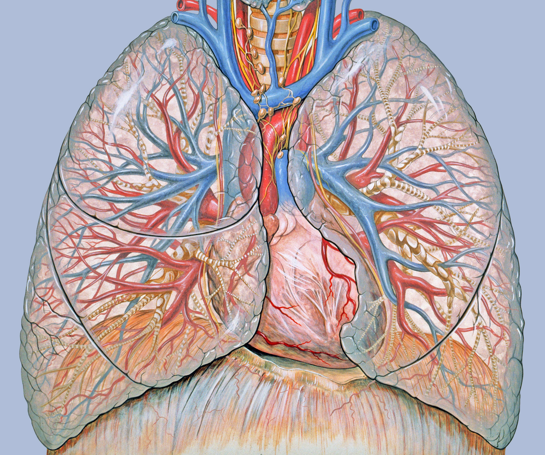
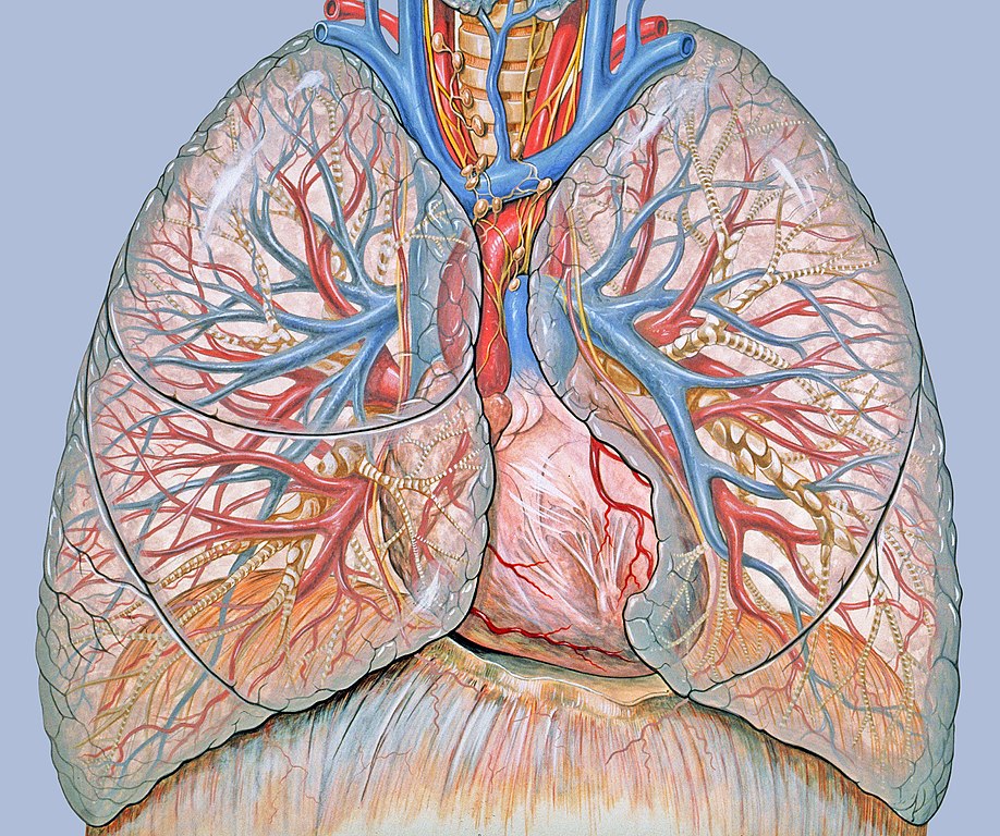
General thoracic anatomy, done specifically to support chest and heart imaging techniques. Generated for multimedia teaching projects by the Yale University School of Medicine, Center for Advanced Instructional Media, 1987-2000. Errors
Keywordschest, lungs, heart, trachea, anatomy, thoracic, thorax, pulmonary, anatomy, cardiac anatomy, diaphragm |
| ތާރީހް | |
| މަސްދަރު | Patrick J. Lynch, medical illustrator |
| Author |
|
| ހުއްދަ (Reusing this file) |
Creative Commons Attribution 2.5 License 2006 |
| Other versions |
 |
Licensing
This file is licensed under the Creative Commons Attribution 2.5 Generic license.
- You are free:
- to share – to copy, distribute and transmit the work
- to remix – to adapt the work
- Under the following conditions:
- attribution – You must give appropriate credit, provide a link to the license, and indicate if changes were made. You may do so in any reasonable manner, but not in any way that suggests the licensor endorses you or your use.
ޞަފްޙާގެ ތާރީޚް
Click on a date/time to view the file as it appeared at that time.
| ތާރީޚް/ގަޑި | ތަމްބްނެއިލް | Dimensions | މެމްބަރު | ޚިޔާލު | |
|---|---|---|---|---|---|
| މިހާރު | 15:15, 11 ޖުލައި 2011 | | 2,304 × 1,927 (1.35 MB) | Materialscientist | removed watermark, cropped |
| 08:25, 23 ޑިސެމްބަރު 2006 |  | 2,640 × 1,927 (1.43 MB) | Patrick.lynch | Patrick J. Lynch; illustrator; C. Carl Jaffe; MD; cardiologist Yale University Center for Advanced Instructional Media Medical Illustrations by Patrick Lynch, generated for multimedia teaching projects by the Yale University School of Medicine, Center for |
ފާލަންތައް
The following 4 pages use this file:
Global file usage
The following other wikis use this file:
- Usage on el.wikipedia.org
- Usage on en.wikipedia.org
- Usage on es.wikipedia.org
- Usage on es.wikibooks.org
- Usage on eu.wikipedia.org
- Usage on fr.wikipedia.org
- Usage on gl.wikipedia.org
- Usage on he.wikipedia.org
- Usage on id.wikipedia.org
- Usage on kbp.wikipedia.org
- Usage on kk.wikipedia.org
- Usage on lb.wiktionary.org
- Usage on pl.wikipedia.org
- Usage on sd.wikipedia.org
- Usage on ta.wikipedia.org
- Usage on uk.wikipedia.org